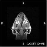
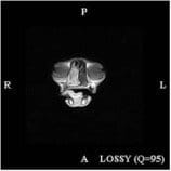
 Mopsy presented to his vet with a history of increased sneezing and occasional nasal discharge which appeared purulent. The primary practitioner was suspicious of nasal neoplasia but could not visualize anything up the nostril or behind the soft palate. Radiographs were equivocal.
 Mopsy went back to his own vet for conservative management of the mass.
These are some of the images obtained from Mopsy’s MRI scan. You can see that there is an obvious soft tissue density within the right nasal chamber. There is deviation of the midline on the transverse image but no evidence of a fluid-gas interface.

