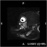
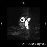
 Petal was a young bouncy Bulldog – her worried owners presented with her as they’d noticed that her eye was bulging out of its socket. The primary veterinary surgeon did a fine needle aspirate and could not aspirate any material – he was concerned that the mass was neoplastic.
 This patient was then referred for MRI to investigate her exophthalmos. MRI is ideally suited to better investigation of this condition and enables diagnosis of the underlying pathology in the majority of cases. The MRI revealed an unusual complex cystic lesion within the orbit resulting in displacement and compression of the globe.
With this information, the primary vet was able to advance a needle to drain the cyst and gave appropriate medical therapy. A repeat scan six weeks later showed that the cyst had resolved. MRI is well suited to orbital disease.

